Pregnancy ultrasound: How many pregnancy scans do you have - and what to expect
Pregnancy ultrasounds take place around 12 and 20 weeks to check on the health of your baby, uterus and placenta.


Parenting advice, hot topics, best buys and family finance tips delivered straight to your inbox.
You are now subscribed
Your newsletter sign-up was successful
A pregnancy ultrasound is a routine scan that looks at your baby while it is growing inside the womb. The NHS offers mothers two or three - usually an early pregnancy scan at 12 weeks, followed by a 20-week and sometimes 36-week scan. Though if there are complications, you're expecting twins or multiple babies, or you have conceived with IVF, you will be recommended more.
To understand how pregnancy ultrasounds work, we spoke to Obstetrician Prof Alexander Heazell, who told us, “Ultrasounds use very high-frequency sound waves. These are emitted from the probe we use on your abdomen and they bounce back to the computer as the sound gets reflected. The probe will measure the sound waves: when the sound waves hit bone, they come back quickly, whereas sound waves hitting fluid come back slower or not at all. The computer then interprets and draws the image for you to interpret.”
How many pregnancy ultrasounds do you have during pregnancy?
The number of pregnancy ultrasounds you'll need can vary. For standard, uncomplicated pregnancies 2-3 scans are recommended by the National Institute of Clinical Excellence. The main ones are commonly known as the “12-week dating scan” and “20-week anomaly” scans. These scans are offered to everyone.
Some trusts have introduced a “presentation scan” at around 36 weeks to check your baby’s position in the womb and where their head is (head down, head up, or “transverse” - sideways). Your midwife will tell you what to expect.
Early pregnancy ultrasound scan
Sometimes, it might be necessary to have an early scan (before 12 weeks), at the early pregnancy unit. These scans are often done in a gynaecology unit specifically equipped for scanning very small structures.
Early pregnancy scans are often recommended if you're when there is a concern you could be having a miscarriage. This includes finding out if you have an ectopic pregnancy - (a pregnancy that has formed somewhere other than your uterus). This can be very dangerous, so it needs to be diagnosed quickly.
What to expect at an early pregnancy ultrasound:
Parenting advice, hot topics, best buys and family finance tips delivered straight to your inbox.
Pregnancy ultrasound scans are usually performed over the abdomen (from the outside of your stomach). However, scans performed before 12 weeks of gestation are often done transvaginally. This means that you will be asked to consent to the sonographer using a probe inside your vagina. This is because the structures they are looking at are very small. This makes them harder to see using a traditional probe on your abdomen.
However, if having a transvaginal scan is something you are not comfortable with, for any reason, discuss this with your healthcare team. You do not have to consent to a transvaginal scan, it is up to you. Before this type of scan, you will be asked to empty your bladder as this makes the scan easier for the sonographer.
What happens at a pregnancy scan?
What happens at each scan depends on the gestation of your pregnancy. What is important is understanding the reason for the scan. Some people will have more scans than others.
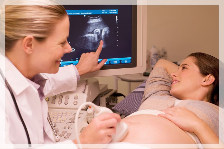
You will usually be asked to lay down on a bed in a darkened room. The sonographer will put some special gel on your abdomen to help the probe make good contact with your skin. It’s sometimes a little cold and it can be quite messy, but it usually washes off clothes easily. The gel increases the effectiveness of the ultrasound waves.
There is a screen, where you can see the sound waves echoed back and translated into a picture. Some of the images might seem confusing as the sonographer does their checks - you need lots of training to correctly understand a scan. However, you will have things explained to you as the sonographer does their checks and they will show you what they are measuring.
You can usually take someone with you, but this might depend on your hospital trust, any Covid restrictions in place and who is doing your scan (private or NHS). Pictures are often provided at NHS scans, but you may have to pay extra for them, depending on your trust.
12 weeks pregnancy scan
The scan done at around 12 weeks is usually the first scan offered by the NHS and is most commonly known as the dating scan. Despite the name, it's actually offered between 11 and 13+6 weeks of pregnancy. This is because as the evidence demonstrates this is the best time for working out when your baby is due. It is also the best time to complete screening for trisomy and chromosomal differences like Down’s Syndrome (also known as chromosomal abnormalities).
The scan is performed to identify :
- When your baby is due (dating).
- Any early issues in development - studies show approximately 50% of structural differences (abnormalities) can be detected in the first trimester.
- Whether you are having more than one baby.
- If you are having two or more babies: what type of twin, triplet or quadruplet pregnancy you are having.
Your baby’s head diameter and crown to rump length (from their head to their bottom) will be measured. These measurements will then be used to calculate your estimated due date.
This might differ from the date calculated from your last menstrual period, but that isn’t usually a cause for concern. This is because you can sometimes ovulate earlier or later than expected according to your period dates.
The NHS will use the result of your dating scan to make recommendations on your care. However, all pregnancy ultrasound scans are optional and you do not have to have them if you do not wish to. Discuss this with your midwife if you have any concerns about ultrasounds, for any reason.
Amanda Bastianelli, Sonographer told us, "It’s important to remember that an ultrasound is a snapshot of the current presentation on that day. Some things will make an ultrasound more difficult. This includes the position of your baby, how much amniotic fluid is present, whether your bladder is full enough and if there’s more than one baby. At the 12-week scan there’s only so much we can check because not everything has developed yet. So it’s not as thorough as the later anomaly scan."
Prepare for your scan by:
- Reading any patient information that is given to you.
- Drinking enough water so you have a full bladder (except transvaginal scans, where you need an empty bladder).
- Taking your pregnancy notes with you.
When does screening for Down's Syndrome happen?
Screening for Down’s Syndrome also takes place at the 12-week scan. This screening is also known as a combined screening because it uses ultrasound scans and the results of blood tests to give you a level of chance that your baby has Downs Syndrome.
The ultrasound scan part of the screening looks at the “nuchal translucency” - the amount of fluid in your baby’s neck. The measurement of the fluid in the neck, combined with the gestational age of your baby, your age, and your blood results will be used in a calculation. This will give you your chance - high or low chance - that your baby has a chromosomal difference.
Source: British Medical Ultrasound Society
This scan is a screening test, meaning that it can estimate the chance of your baby having Down's, but it can't diagnose. If the result comes back and shows that it's likely, you might then decide to have a diagnostic test such as amniocentesis or chorionic villus sampling to confirm.
A blood test combined with your scan can help identify how likely it is that your baby has Downs syndrome, as well as Edwards Syndrome or Pateaus Syndrome.
Screening is completely optional and some people decide not to accept it. Others decide to have some of the screening tests and not others. Speak to your midwife if you are unsure about whether to accept screening and need further, personalised advice about what to decline and accept.
If the results of your screening tests come back as “high chance”, that does not necessarily mean that your baby definitely has a chromosomal difference.
Additional tests will be offered to you and you will receive lots of information about the next steps. You may decide you do not want these tests and are happy to proceed with your pregnancy without them.
It can be very anxiety-inducing to receive your results and quite overwhelming to understand all the information given to you. You might not know how you feel about your results, especially if it is later confirmed that your baby has a chromosomal difference.
Further support is available from:
- Antenatal Results and Choices
- Positive about Down Syndrome
- Support Organisation For Trisomy 13/18
- Down’s Syndrome Association
Measuring your baby’s growth
During your ultrasound, you'll notice the sonographer is drawing a line across the screen to take your baby's measurements. Prof Alex Heazell, Obstetrician told us, "We mostly take 3-4 measurements to give us our estimation of growth and fetal weight. We take two head measurements: across the skull, above the ears, and also around the head, like the brim of a hat. We also measure around the tummy and the femur (thigh bone) length. We use those measurements in a formula that comes up with an estimate. Like any formula, it’s not perfect and there is usually a variance of about 10%, which of course can be quite significant. The bigger the baby is, the bigger the number of grams that could be different. In general, we are better at picking up small babies than big babies."
These prenatal growth scans are a useful indicator of how your baby is doing within the womb. However, they must be taken as a part of the “puzzle” making up how your pregnancy is progressing, rather than an indication on their own. All babies and all pregnancies are different and some people have smaller babies than others.
Research shows, Intrauterine growth restriction (a baby who is measuring small for dates) happens in around 10% of pregnancies. Their accuracy is not 100%. In fact, studies show that detecting overly small and overly large babies can be difficult.
Your midwife will also offer you “fundal height measurements” from 25 weeks, which measure the height of your bump with a tape measure. This tells them how your baby is growing. Sometimes, if your uterus seems larger or smaller than most other pregnancies at the same gestation, this might mean there is a problem. However, it’s important not to worry as everyone is different. Everyone carries their baby differently and usually a scan will show your baby is growing exactly as he or she should.
What happens if you receive worrying news at your ultrasound?
Unfortunately, you may discover that you have miscarried your baby at the 12-week scan. This can be incredibly traumatic and upsetting. If this happens, support is available from the healthcare team providing the scan. The next steps will be discussed with you and you will usually have an opportunity to return home and make a decision about what to do next.
Support is available from your GP, midwife, and from various charities:
- The Miscarriage Association
- The Worst Girl Gang Ever community and podcast
20 weeks pregnancy scan
The 20-week pregnancy scan can be performed between 18 and 20+6 weeks of pregnancy and is often known as the anomaly scan.
The scan is performed to identify :
- The bones of all four limbs and fingers/toes.
- All four chambers of the heart.
- Structure of the brain.
- Their spinal cord.
- Their face, nasal bone and mouth (checking for cleft lip).
- Kidneys (that both are present and functioning as expected).
- The shape, size and function of their stomach and abdomen.
These examinations will usually reveal any major differences in your baby (abnormalities) but will not rule them out entirely. Your results will be explained to you. If any measurements are not able to be done, you may be offered another scan.
The scan may take up to 30 minutes, but will obviously be longer if there are issues with getting good images, or if you are expecting more than one baby.
Sometimes, there are problems with looking at the baby if it is not in a good position. You might be asked to go for a walk, have something to eat and drink and come back that same day to try to get your baby to move to a better position. Sometimes you might need to attend another, new appointment.
Prepare for your scan by:
- Check and follow the patient information given to you.
- Make sure you attend with a full bladder.
- Don’t forget to take your pregnancy notes to your appointment.
When will I find out the sex of my baby?
Many people think that the purpose of the 20-week scan is finding out the sex of your baby. However, not all trusts will tell you at your 20-week scan. It depends on the policy of your hospital.
The 20-week scan is usually the earliest that an NHS sonographer will offer an opinion on what genitals your baby has. This opinion cannot be guaranteed but is usually found to be correct at birth.
Not everyone wants to know during pregnancy and would prefer to find out when their baby is born. Everyone is different and your decision is personal. Check with your partner about what they would prefer and discuss it together before you attend your 20-week scan.
Some private scanning clinics offer paid ultrasounds to tell you the sex of your baby (usually advertised as gender scans). These are offered at different times depending on the policy of the clinic. Some clinics will scan and offer an opinion on your baby’s sex as early as 14 weeks.
If you get an early ultrasound before 12 weeks, there are some people who say that by using either the Ramzi Theory or Nub Theory, you can tell the gender of your baby but there's no scientific evidence to back it up.
What happens if there is an issue at my 20 week anomaly scan?
There are many things that can be identified at the 20-week anomaly scan and your results should be explained to you in detail. Studies show structural differences (referred to as abnormalities) are present in about 3% of pregnancies.
If there are any signs of structural differences (abnormalities) picked up in the scan, you will be taken to a private room and have the results discussed with you.
You may be offered more diagnostic tests and further scans, depending on what has been found. Not all of the initial results will mean that your baby will be affected - sometimes these results are for conditions that can resolve themselves in the pregnancy. However, some results might mean that you need to consider some next steps. These can include further tests or conversations about whether you wish to continue with your pregnancy.
Support after pregnancy ultrasound results
You will be supported to understand exactly what your scan results mean and given information to help you make decisions about what tests to undergo next. It’s likely you will be offered an appointment with a Fetal Medicine Doctor to understand more about what has been identified.
These decisions are sometimes tough and there is not always a “right” or “wrong” decision. Depending on their results, some people decide to undergo a termination for medical reasons (TFMR). Some people call this “compassionate induction”.
However, others take the decision not to do so and continue the pregnancy. It really depends on the condition that’s identified, how it impacts the child, your feelings and your personal beliefs as to what you decide to do.
These are not easy decisions to make. Whatever you decide, you will need support and maybe even counselling to deal with the resulting feelings.
Support for those considering termination for medical reasons:
- Baby Loss Awareness Alliance Members
- SANDS
- Antenatal Results and Choices
- Tommy’s Charity
- The Worst Girl Gang Ever
Support networks for continuing your pregnancy after antenatal diagnosis:
Where to find support for continuing your pregnancy after antenatal diagnosis will depend on what condition or difference/disability your baby has. There is a range of charities out there that can help you understand the diagnosis and what kind of support you and your child might need:
- Genetic Alliance for rare or genetic conditions
- Positive about Down Syndrome
- Carers.org
- Sandcastle Trust
- Action for Children
- The UK government’s advice and support
- Mencap
Other pregnancy ultrasounds
During your pregnancy, you might be recommended to have extra scans to check your baby, the placenta or both. There are many different reasons that extra pregnancy ultrasounds might be recommended. Those expecting more than one baby, or who have a health problem that might affect their pregnancy (such as diabetes), will have more regular growth scans.
Two of the most common reasons for extra pregnancy ultrasounds are:
- If your uterus or baby is not growing as expected
- If you have a problem with your placenta
Can ultrasounds harm my baby?
The safety of ultrasound scans is quite well established in medicine, as there is not any evidence that they are unsafe. Many studies have been performed to determine whether there are any dangers and so far, there is little evidence that they cause harm. Millions of pregnancies have been scanned since ultrasounds were introduced as standard in every country across the world and no adverse effects have been discovered, as yet.
There are some minimal concerns about heating of the tissues and bones of early developing fetuses, however. Studies on this are limited but indicate that it is not a cause for concern where exposure is limited in time and intensity.
NHS Sonographers use the “ALARA” principle: “as low as reasonably possible” and this refers to using low-powered ultrasound sound waves, for as short a time as possible, only when necessary.
A World Health Organisation review of the literature (examining data from 41 studies) concluded “According to the available evidence, exposure to diagnostic ultrasonography during pregnancy appears to be safe”

Professor Alexander Heazell, Senior Clinical Lecturer in Obstetrics and Clinical Director of the Rainbow Clinic at Tommy’s Research Centre, University of Manchester.

Amanda Bastianelli is a specialist midwife sonographer with the NHS and at Expectancy Scanning Studios Ltd in Kent. With over 20 years of experience, she completed a Diploma in Midwifery at the University of Greenwich, before gaining a Postgraduate Certificate in Medical Imaging, Obstetrics & Gynaecology at Canterbury Christ Church University.

Tannice Hemming has worked alongside her local NHS in Kent and Medway since she became a parent and is now a mum of three. As a Maternity Voices Partnership Chair, she bridged the gap between service users (birthing women and people, plus their families) and clinicians, to co-produce improvements in Maternity care. She has also worked as a breastfeeding peer supporter. After founding the Keep Kent Breastfeeding campaign, she regularly appears on KMTV, giving her views and advice on subjects as varied as vaccinations, infant feeding and current affairs affecting families. Two of her proudest achievements include Co-authoring Health Education England’s E-learning on Trauma Informed Care and the Kent and Medway Bump, Birth and Beyond maternity website.